Overview
The spine (vertebral column, spinal column) is a complex entity of interconnecting bones (vertebrae), mobile joints, nerves, ligaments and muscles all working together to provide support, stability and flexibility, especially on extending, bending and rotation. The vertebral column usually consists of 33 vertebrae, arranged in five regions: cervical, thoracic, lumbar, sacral and coccygeal.
Anatomy (Cervical region)
The cervical spine consists of seven vertebrae, which are the smallest and uppermost in location within the spinal column and supported by strong ligaments and several muscles. The cervical vertebrae connect the neck to the thoracic spine (the upper back). All seven vertebrae are numbered. C1, the first vertebra in the column (closest to the skull), is also known as the atlas. C2, the vertebra below it, is also known as the axis. C stands for cervical.
The cervical spine serves several crucial functions, including: 1. Supporting the head and its movements, 2. Protecting the spinal cord and the spinal nerves, 3. Facilitating blood flow to the brain (vertebral openings or foramen in the cervical spine provide a passage for vertebral arteries to ensure proper blood flow to the brain).
The Atlanto-Occipital joint is where the lower part of the occipital bone (base of the skull) connects with the first cervical vertebra, called the atlas. This region facilitates the passage of the spinal cord out of the skull and down into the cervical spine.
Anatomy (Thoracic region)
The thoracic spine is located in the upper back and is the longest region of the spine. The thoracic spine consists of twelve vertebrae, labelled from T1 down to T12. T stands for thoracic. T1, the top thoracic vertebra, connects with the lowest cervical vertebra, C7, in the cervical spine above, while the lowest thoracic vertebra, T12, with the first lumbar vertebra, L1, in the lumbar spine below. Additionally, these vertebrae serve as attachment points for the ribcage.
The thoracic spine has several crucial functions, including: 1. Protecting the spinal cord and the spinal nerves, 2. Anchor the rib cage (the rib cage forms a bony structure to surround and protect internal organs, such as the lungs and the heart).
The costovertebral joints are where the thoracic vertebrae connect or articulate with the ribs.
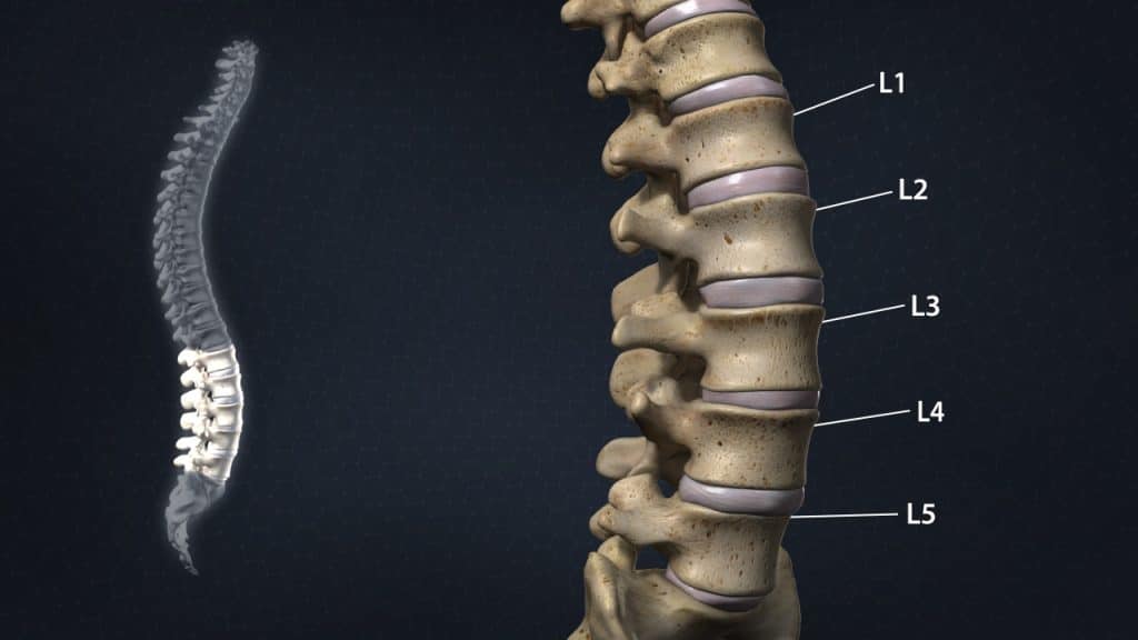
Anatomy (Lumbar region)
The lumbar spine consists of five vertebrae which connect the sacrum (triangular bone at the bottom of the spine) to the thoracic spine (the upper back). All five vertebrae are numbered. L1, the first lumbar vertebra, is closest to the thoracic spine. L5, the lowest lumbar vertebra, is closest to the sacrum. L stands for lumbar.
Anatomy (Sacral region)
The sacral region is composed of five fused sacral vertebrae, labelled S1 to S5. These vertebrae form a solid mass of triangular bone at the bottom of the spine, referred to as the sacrum. The sacrum provides strength and stability to the pelvis.
The sacroiliac joints are the joints between the sacrum and the iliac bones (bones of the pelvis), which are connected by strong ligaments. They are essential for effectively transmitting the weight of most of the body to the pelvis and the lower extremities. Movement of the sacroiliac joints is restricted. Small movements at these joints serve shock absorption and forward/backward bending.
Anatomy (Coccygeal region)
The coccygeal region, known as tailbone, is composed of four fused coccygeal vertebrae. These vertebrae form the coccyx. It provides attachments for various muscles, tendons and ligaments.
Anatomy (A typical lumbar vertebra)
A typical lumbar vertebra (bone of the spine) is composed of two parts. The vertebral body in the front (anterior) and the vertebral arch in the rear (posterior).
The vertebral arch encloses the vertebral foramen (opening). It consists of a pair of pedicles and a pair of laminae. At the junction of the pedicle and lamina on the right and left side, bony protrusions project upward and downward, known as superior and inferior articular processes.
The pars interarticularis (bridge between two bones) connects the superior and inferior articular processes. The pars interarticularis may also be referred to as the isthmus.
At the mid-point of the vertebral arch, a bony protrusion known as spinous process projects backward and downward. On either side of the spinous process, there are bony protrusions known as transverse processes. Similar to the spinous process, these processes provide attachments for ligaments and muscles.
The vertebrae (bones of the spine) articulate with one another at joints between their bodies and between their articular processes. The intervertebral discs provide the strongest attachment between the bodies of the vertebrae. In addition to the discs, the bodies are united by strong supporting ligaments.
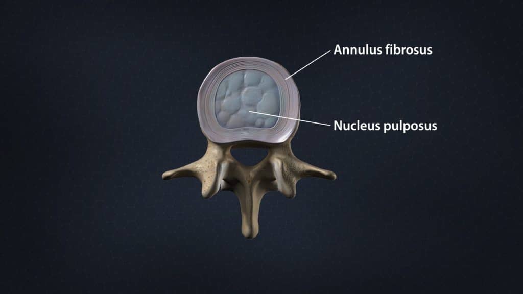
Anatomy (Intervertebral discs)
The intervertebral discs are shock absorbers that are located between the bones of the spine, called vertebrae (hence the name intervertebral). They are designed to help the back stay flexible, while resisting forces and to allow bending, flexion and twisting of the spine. A healthy, well-hydrated disc contains a great amount of fluid in its centre, known as the nucleus pulposus, which provides cushioning and flexibility to the spine.
Anatomy (Facet joints)
The facet joints or zygapophyseal joints are a set of joints between the articular processes of two adjacent vertebrae. They are covered with cartilage and are surrounded by a lubricating capsule that enables the vertebrae to bend and twist. Each capsule has a rich supply of tiny nociceptive nerve fibers and implicates this structure as a potential source of pain. Similar to other joints in the body, these joints are vulnerable to inflammation and degeneration.
The facet joints provide support, stability and mobility to the vertebrae, especially on extending, bending and rotation. Furthermore, the articulation between the superior (above) and inferior (below) articular facets at the zygapophyseal joints as well as the intervertebral discs play an important role to prevent a vertebra slip over the adjacent one.
Anatomy (Spinal Nerves)
The entire length of the vertebral column has a large central canal or passage, called the spinal or vertebral canal. In the cervical and thoracic regions of the spine, the spinal cord descends through this space. In the lumbar region of the spine, this space contains a bundle of nerve roots, known as cauda equina. Openings or holes to each side of the canal, called the neural foramen (intervertebral foramen – foramen between the two adjacent vertebrae) provide pathways for the nerve roots that travel from the spine to other parts of the body.
Reference
- Moore K.: Clinically Oriented Anatomy. Third Edition. Williams & Wilkins 1992.